Visual Enabling for Precision Surgery
by Klaus Kansy
The project 'Visual Enabling for Precision Surgery' (VEP) will develop a computer-based 3D image guidance system for neurosurgical interventions which are supported by an open magnetic resonance imaging (MRI) system from General Electric. Open MRI systems can generate intra-operative images which give important feedback for the precise positioning of surgical devices.Image-guided surgery is a new medical discipline where different imaging modalities like computed tomography (CT), MRI, ultrasound, etc. are used not only for diagnosis but also for guiding a surgeon during the intervention. For example, a physician can localize a brain tumor in MRI and plan the best therapy and access way. Then, a computer system can be used to precisely position the surgical instruments during the intervention. For interventions into soft tissues like brain, geometrical data gathered from preoperative images are corrupted by the intervention and have to be updated by real-time measurements. Open MRI system provide such data during the intervention.
The surgeon needs good spatial and structural orientation to perform his difficult task which is not readily provided by an unrelated set of images of different quality and content. Therefore, the focal element of the VEP project is a 3D reference scene realized as an augmented reality system combining various types of medical data sets and synthetic images. The physician will experience this surgical orientation environment (visual enabling system) as a kind of interactive graphical 3D scene showing all relevant data for the intended intervention. The design approach requires close interdisciplinary co-operation among computer scientists, medical physicists, and human factors specialists with radiologists, surgeons, and other medical experts.
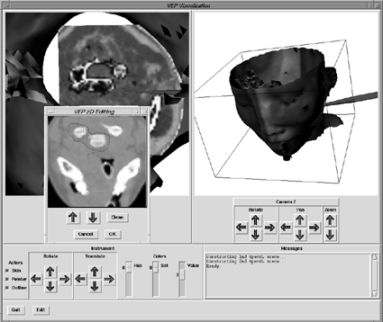
Figure: VEP window showing head with MRI cut, an survey image with surgical device, and an editing window for interactively locating a tumor.Technically, 3D and 2D data sets of various available imaging sources have to be registered (ie, geometrically aligned) and integrated. During the intervention, real-time measurements will update and complement these data. Synthetic images of surgical devices and the planned access will enhance the medical data. The system will be implemented using OpenGL on workstations ranging from SGI Onyx down to PCs.
The VEP system is an example of a medical enabling system which can be attached to new medical high-tech systems in order to make them intuitively accessible for the average physician. Further information on the web at: http://zeus.gmd.de/hci/projects/vep/vep.html
Please contact:
Gernoth Grunst - GMD
Tel: +49 2241 14 2346
E-mail: grunst@gmd.de